Calibration for Clinical Recording of Periodontal Status
The goal of recording a periodontal status is to record recession, probing depth, and attachment level at six points per tooth or implant throughout the dentition with millimeter precision. For all measurements performed with the periodontal probe, the values read on the probe are rounded up.
1. Gingival Margin, Probing Depth and Attachment Level
At each site, the "Gingival Margin" value is measured first, followed immediately by the "Probing Depth" value. The "Attachment Level" value is automatically calculated in the online periodontal status and displayed graphically with a blue line.
The first value "Gingival Margin" is the millimeter distance from the clinical gingival margin to a reference point, such as the cemento-enamel junction. If an existing crown or filling margin is no more than 3mm apical to the original cemento-enamel junction, these margins are used as reference points. Otherwise, a virtual reference point at the level of the original cemento-enamel junction is chosen.
The second value "Probing Depth" is the millimeter distance from the clinical gingival margin to the bottom of the gingival sulcus or periodontal pocket.
The third value "Attachment Level" is calculated by the online periodontal status for each site according to the following formula:
Attachment Level (mm) = Probing Depth (mm) – Gingival Margin (mm)
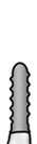
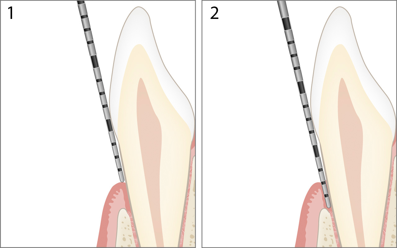
Healthy Periodontium
In the healthy periodontium, the cemento-enamel junction lies below the gingival margin and immediately above the attachment level (no attachment loss).
The values for "Gingival Margin" 1 and "Probing Depth" 2 are identical in this case.
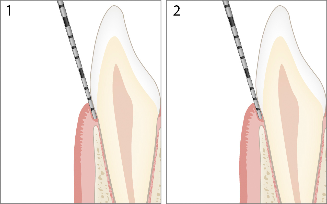
The calculation of the attachment level in the figure shown above is:
Attachment level 0mm = Probing depth 2mm – Gingival margin 2mm
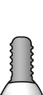
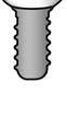
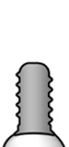
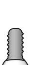
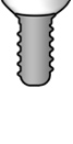
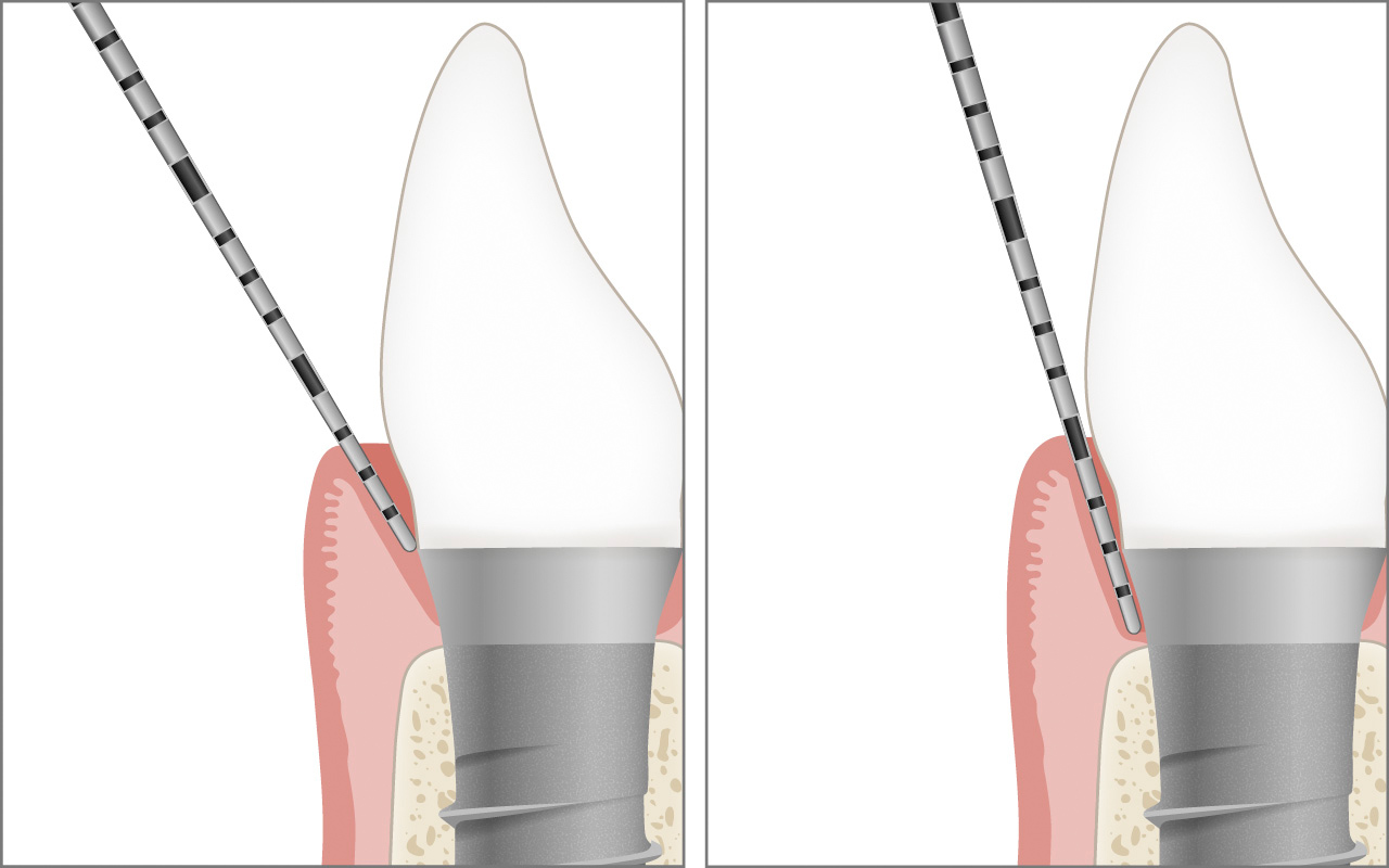
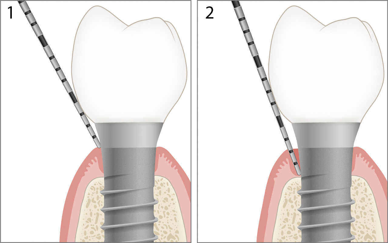
Healthy Peri-Implant Tissue
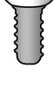
In healthy peri-implant tissue, the margin of the suprastructure lies slightly below the peri-implant mucosa (no alveolar bone loss).

The calculation of the alveolar bone level (attachment level) in the figure shown above is:
Alveolar bone level 2mm = Probing depth 3mm – Gingival margin 1mm
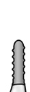
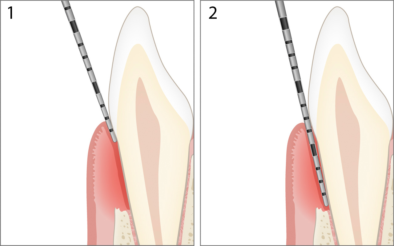
Hyperplastic Periodontium (Overgrowth)
In the hyperplastic periodontium, the cemento-enamel junction may lie far below the gingival margin (>3mm), but still immediately above the attachment level (no attachment loss).
The values for "Gingival Margin" 1 and "Probing Depth" 2 are also identical in this case.

The calculation of the attachment level in the figure shown above is:
Attachment level 0mm = Probing depth 5mm – Gingival margin 5mm
Note: Pockets with probing depths of 4mm or more without attachment loss are referred to as pseudo-pockets.
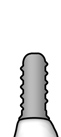
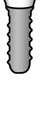
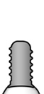
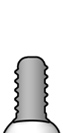
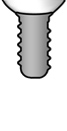
Healthy Peri-Implant Tissue in the Aesthetic Zone
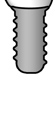
In healthy peri-implant tissue in the aesthetic zone, the margin of the suprastructure lies further below the peri-implant mucosa (no alveolar bone loss).
The calculation of the alveolar bone level (attachment level) in the figure shown above is:
Alveolar bone level 2mm = Probing depth 5mm – Gingival margin 3mm
Periodontal Pocket
In the diseased periodontium, the cemento-enamel junction may lie above or below the gingival margin. The distance from the gingival margin to the bottom of the periodontal pocket is recorded as the probing depth 2.
The calculation of the attachment level in the figure shown above is:
Attachment level 5mm = Probing depth 7mm – Gingival margin 2mm
Note: Pockets of 4mm or more that remain after periodontal therapy is completed are called residual pockets.
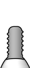
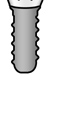
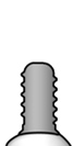
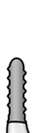
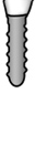
Peri-Implant Pocket
In peri-implant diseased tissue with alveolar bone loss, the margin of the suprastructure may lie slightly below or above the peri-implant mucosa. The distance from the margin of the peri-implant mucosa to the bottom of the peri-implant pocket is recorded as the probing depth 2.

The calculation of the alveolar bone level (attachment level) in the figure shown above is:
Alveolar bone level 6mm = Probing depth 7mm – Gingival margin 1mm
Gingival Recession
In the case of gingival recession, the gingival margin lies apical to the cemento-enamel junction. The measured value for "Gingival Margin" 1 is then given with a negative sign.
The calculation of the attachment level in the figure shown above is:
Attachment level 6mm = Probing depth 2mm – Gingival margin -4mm
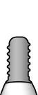
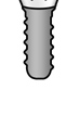
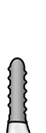
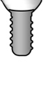
Peri-Implant Recession
In the case of peri-implant recession, the margin of the peri-implant mucosa lies apical to the margin of the suprastructure. The measured value for "Gingival Margin" 1 is then given with a negative sign.
The calculation of the alveolar bone level (attachment level) in the figure shown above is:
Alveolar bone level 4mm = Probing depth 2mm – Margo Mucosae (Gingivae) -2mm
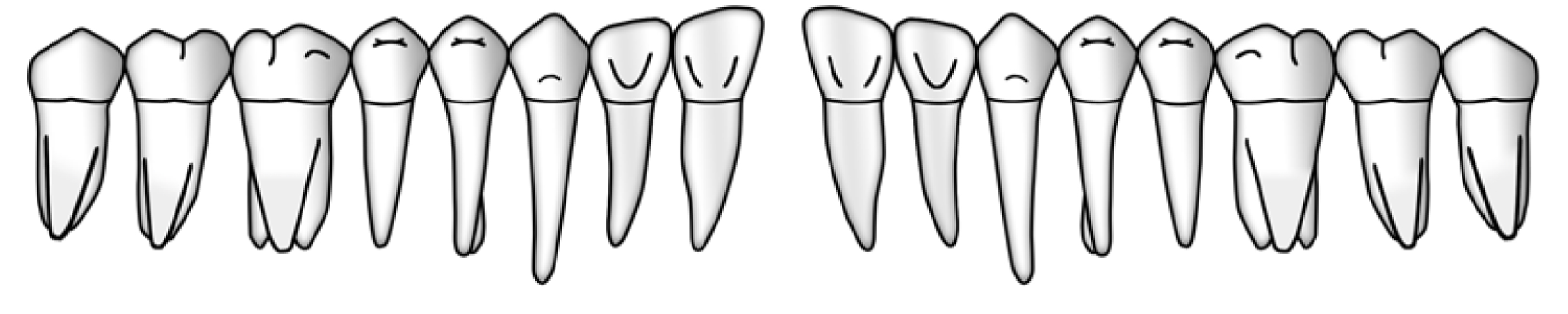
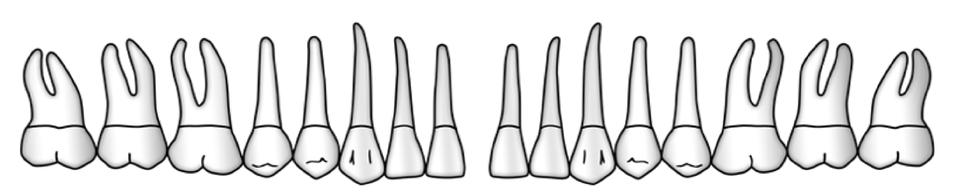
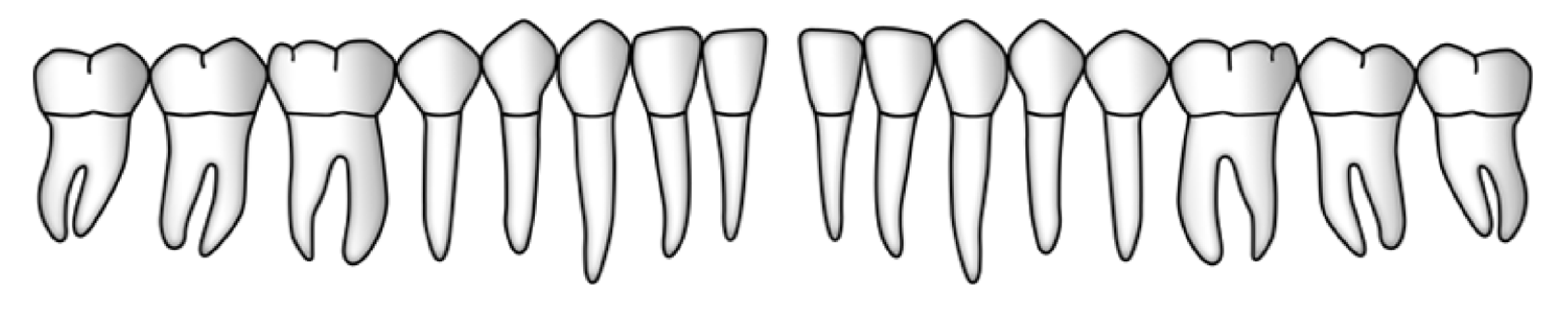
Six Recording Sites per Tooth or Implant
Crucial for the recording of all periodontal or peri-implant measurements is the choice of the correct six sites around the tooth or implant. For this purpose, the tooth or implant is viewed from the occlusal aspect and divided into 6 sections. For each of these sections, the site with the highest probing value is determined and measured.

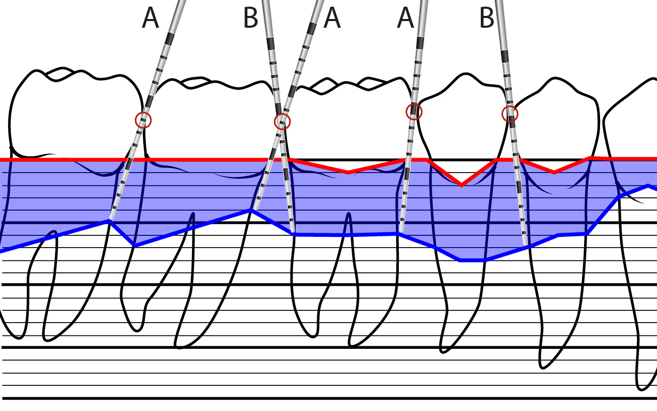
Angulation of the Periodontal Probe
When measuring probing depth, the probe is guided apically along the tooth surface. It can tilt in the mesio-distal axis A or B, while the bucco-oral direction must be held parallel to the tooth longitudinal axis.
2. Furcation Involvement
The furcations of the molars and the first premolars in the upper jaw are examined with a furcation probe. The horizontal component of the total penetration depth is divided into grades 0 - 3 according to the following criteria (Hamp et al., 1975).

Grade 0 Furcation entrance not detectable
Grade 1 Furcation detectable, horizontal probing value ≤3mm
Grade 2 Furcation detectable, horizontal probing value >3mm
Grade 3 Furcation open through and through
3. Tooth Mobility
The tooth mobility of all existing teeth is measured bidigitally and divided into grades 0 – 3 according to the following criteria (Miller, 1950).
Grade 0 Physiological mobility
Grade 1 Increased mobility up to 1mm horizontally
Grade 2 Increased mobility more than 1mm horizontally
Grade 3 Severe mobility in a vertical direction
Literature
Miller S. C., Textbook of Periodontia, 3rd edition, The Blakiston Co., Philadelphia and Toronto, 1950.
Hamp S. E., Nyman S., Lindhe J., Periodontal treatment of multirooted teeth. Results after 5 years. J. Clin. Periodontol. 1975;2:126–135. doi: 10.1111/j.1600-051X.1975.tb01734.x.


 English (UK)
  English (US)
  German
  French
  Italian
  Español
  Basque
  Português (BR)
  Polish
  Danish
  Romanian
  Bulgarian
  Croatian
  Slovenian
  Chinese
  Japanese
  Russian
  Georgian
  Azerbaijani
  Turkish
  Lithuanian
  Malaysian
  Vietnamese
  Dutch
Dutch  Hungarian
  Czech
Czech  Greek
Greek  Finnish
  Swedish
  Norwegian
  Ukrainian